Beta-galactosidase as indicator of senescent and cancer cells

Yesterday I was listening to a news segment on CBC Links to an external site.about cervical cancer screening results that currently have 6-month delays in parts of Canada due to post-pandemic backup and staff shortages. Coincidently, while researching beta-galactosidase, I discovered that this enzyme is an important biomarker for certain cancers, including ovarian cancer (Fan et al., 2021) and, most recently, has been found to be over-expressed in gastric cancer as well (Kubo et al., 2021). 

Human beta-galactosidase is highly expressed in senescent cells, which are cells that stop dividing but continue to exist and metabolize. Accumulation of senescent cells is an indicator of aging in general, and the presence of these cells is associated with a risk for many diseases including cancer, osteoarthritis, atherosclerosis (arterial plaque build-up), Alzheimer’s, diabetes, and more (Childs et al., 2017) (Chen et al., 2022). Interestingly, cellular senescence seems to be one of the body’s defenses against tumor growth, in that it’s a way for the body to stop the division of cells that have shortened telomeres and may have mutations (Campisi, 2001). Yet, secretions by senescent cells also cause disease (Childs et al., 2017) and even a small number of senescent cell secretions such as cytokines can have a significant impact on cells throughout the entire body and cause or increase disease (Burton, 2009).  And so, the presence of an accumulation of human beta-galactosidase in tissues indicates aging and the onset or development of disease.  

Early cancer detection greatly affects survival rates. Moreover, distinguishing the exact type of cancer aids in treatment decisions. Finding biomarkers that indicate early cancer growth and specify the type of cancer is key to increasing survivorship. Beta-galactosidase is shown to be present in ovarian cancer and most recently associated with gastric cancer (Kubo et al., 2021). Detection of beta-galactosidase has been historically done by a technology called X-gal but has certain disadvantages including that it can only be done in cells that are non-living and the staining takes a long time and is difficult to visualize. However, a new technology called Cellular Senescence Detection Kit - SPiDER-βGal is more sensitive, easier to see, and can be done on live cells (in vivo) (Cellular Senescence Detection Kit - SPiDER-βGal, n.d.).

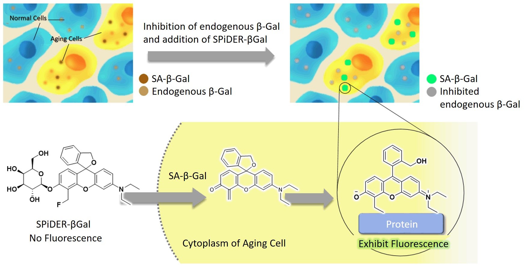
The image shows beta-galactosidase cells illuminated with the application of SPiDER-βGal technology. Source: (Cellular Senescence Detection Kit - SPiDER-βGal, n.d.).

Research has shown that beta-galactosidase fluorescent illumination is a reliable indicator for gastric cancer, liver, and other cancers. B-galactosidase is found in higher concentrations in tumor tissues compared to healthy cells. The presence of beta-galactosidase can be seen visually, with a stronger fluorescence indicating higher concentrations.  Furthermore, the detection is fast and can be seen only 10 minutes after application. This technology could thus be used during surgery, to help surgeons see cancer cells during surgery, namely fluorescence-guided surgery (Ogawa et al., 2021). Also, it can shine for hours, is non-toxic, and will illuminate even at low concentrations. Detecting and removing even small amounts of cancer cells can greatly decrease reoccurrences or metastasis. 

I became curious to learn exactly how the probe lights up beta-galactosidase and it took hours of research to find some semblance of an explanation. I finally came upon J. Zhang et al., the researchers who originally developed the technology and published their findings, in 2017. The probe is a substrate for Beta-galactosidase and ‘turns on’ in a solution with H2O. It can detect even a small concentration of 0.1 nM and has a very high bonding affinity. The probe can distinguish between endogenous Beta-galactosidase cells and non-endogenous. It has a hemicyanine structure (meaning dyes with two aromatic groups) with a D-galactose residue using a glycosidic bond and s fluorescent intensity at 703nm (J. Zhang et al., 2017). 

I will admit I am still not satisfied with this information and want to really understand how this probe lights up the specific substrate groups. Here is an image that I partially understand. I can see that Gal-Pro is the probe. It meets with Beta-galactosidase. The top sugar (chair-shaped) comes off and binds to beta-galactosidase. I have many remaining questions, such as why it bonds and how it illuminates. I am sure I am limited in my understanding as my organic chemistry knowledge is not comprehensive. Perhaps someone can comment and explain what happens on a substrate level and what causes the fluorescence. 

Image from Qiu et al., 2020.

In summary, beta-galactosidase is present in high concentrations in senescent cells and is an indicator of ovarian and gastric cancer. SPiDER-βGal probe is a powerful new technology that can be used for fluorescence-guided surgery to find and remove cancer cells. It can also be used as a fast biomarker in vivo for the presence of gastric and ovarian cancer, even in small concentrations.


REFERENCES


Kubo, H., Murayama, Y., Ogawa, S., Matsumoto, T., Yubakami, M., Ohashi, T., Kubota, T., Okamoto, K., Kamiya, M., Urano, Y., & Otsuji, E. (2021). β-Galactosidase is a target enzyme for detecting peritoneal metastasis of gastric cancer. Scientific Reports11(1), 10664. https://doi.org/10.1038/s41598-021-88982-2Links to an external site.

Fan, F., Zhang, L., Zhou, X., Mu, F., & Shi, G. (2021). A sensitive fluorescent probe for β-galactosidase activity detection and application in ovarian tumor imaging. Journal of Materials Chemistry. B, Materials for Biology and Medicine9(1), 170–175. https://doi.org/10.1039/d0tb02269aLinks to an external site.

Childs, B. G., Gluscevic, M., Baker, D. J., Laberge, R.-M., Marquess, D., Dananberg, J., & van Deursen, J. M. (2017). Senescent cells: an merging target for diseases of ageing. Nature Reviews. Drug Discovery16(10), 718–735. https://doi.org/10.1038/nrd.2017.116Links to an external site.

Chen, Y.-H., Zhang, X., Ko, K.-Y., Hsueh, M.-F., & Kraus, V. B. (2022). CBX4 regulates replicative senescence of WI-38 fibroblasts. Oxidative Medicine and Cellular Longevity2022, 5503575. https://doi.org/10.1155/2022/5503575

Childs, B. G., Gluscevic, M., Baker, D. J., Laberge, R.-M., Marquess, D., Dananberg, J., & van Deursen, J. M. (2017). Senescent cells: an emerging target for diseases of ageing. Nature Reviews. Drug Discovery16(10), 718–735. https://doi.org/10.1038/nrd.2017.116Links to an external site.

Campisi, J. (2001). Cellular senescence as a tumor-suppressor mechanism. Trends in Cell Biology11(11), S27-31. https://doi.org/10.1016/s0962-8924(01)02151-1

Burton, D. G. A. (2009). Cellular senescence, ageing and disease. Age (Dordrecht, Netherlands)31(1), 1–9. https://doi.org/10.1007/s11357-008-9075-y

Cellular Senescence Detection Kit - SPiDER-βGal. (n.d.). Dojindo.eu.com. Retrieved December 8, 2022, from https://www.dojindo.eu.com/store/p/895-Cellular-Senescence-Detection-Kit-SPiDER-Gal.aspx

Ogawa, S., Kubo, H., Murayama, Y., Kubota, T., Yubakami, M., Matsumoto, T., Yamamoto, Y., Morimura, R., Ikoma, H., Okamoto, K., Kamiya, M., Urano, Y., & Otsuji, E. (2021). Rapid fluorescence imaging of human hepatocellular carcinoma using the β-galactosidase-activatable fluorescence probe SPiDER-βGal. Scientific Reports11(1), 17946. https://doi.org/10.1038/s41598-021-97073-1

Zhang, J., Li, C., Dutta, C., Fang, M., Zhang, S., Tiwari, A., Werner, T., Luo, F.-T., & Liu, H. (2017). A novel near-infrared fluorescent probe for sensitive detection of β-galactosidase in living cells. Analytica Chimica Acta968, 97–104. https://doi.org/10.1016/j.aca.2017.02.039

Qiu, W., Li, X., Shi, D., Li, X., Gao, Y., Li, J., Mao, F., Guo, Y., & Li, J. (2020). A rapid-response near-infrared fluorescent probe with a large Stokes shift for senescence-associated β-galactosidase activity detection and imaging of senescent cells. Dyes and Pigments: An International Journal182(108657), 108657. https://doi.org/10.1016/j.dyepig.2020.108657

What are epigenetics and is it a good or bad thing?

This is in response to this question:

What role does DNA mismatch repair have in cancers? According to the slides this week, "Base-excision repair corrects the most common point mutation in humans, the deamination of methylcytosine to thymine." If this repair does not work, is there an abundant amount of genetic information and does that lead to cancer?

Your question really opens Pandora’s box. The deamination of cytosine into uracil or thymine is a hot topic in epigenetic research. Epigenetics is the theory that environment and experiences change the expression of genes, altering phenotypic expression in somatic and even germ cells. I found a helpful Harvard infographic that quickly explains epigenetics, which I thought might be helpful.

(What Is Epigenetics? The Answer to the Nature Vs. Nurture Debate, n.d.)  

As you can see from the infographic, the original concept of DNA was that it was a blueprint that dictates phenotypic expressions. However, scientists now perceive that there are many regulators and modifiers of DNA that contribute to a diversity of gene expression. Additionally, these modifiers can be passed on from generation to generation.

Studies with rats have shown transgenerational modifications of gene expression based on early maternal-infant relations. For example, increased DNA methylation in the prefrontal cortex that causes depressive behavior is observed in rats abused by their mothers. This methylation is then passed on in subsequent generations and observed in male sperm and female/male somatic cells, along with observed depressive behaviors (Gudsnuk & Champagne, 2012). This methylation was not present in the DNA but is now present as an epigenetic marker influencing gene expression and is then passed on in germ cells. In other words, if a mother rat abuses her baby, it causes methylation in the cortex that causes depression in her baby and her baby’s babies. Epigenetics has a particular influence on the brain due to the plasticity of cerebral neurons.

And so, cytosine methylation into thymine (or uracil) is one of the key players in epigenetics. The transition mutation from cytosine to thymine has many implications when not corrected by base-excision repair, from increased variation in immunological defense to oncogenesis.

In terms of immunity, it is crucial to have antibodies that can recognize and defend against a large and ever-evolving diversity of pathogens. Deamination of cytosine is important for the hypermutation of somatic B cells that produce the necessary antibody diversity. Enzymes such as cytosine deaminases are crucial for an efficient immune system and shown to be lacking in humans with hyper-IgM immunodeficiency. Thus, the deamination of cytosine enables B cells to create the immunoglobin diversity necessary for efficient immunity.  (Chahwan et al. 2012).

Counter to that, deamination mutations of cytosine are also implicated in many cancers. Studies have found that activation-induced cytidine deaminases (AID) cause early stop codons in tumor-suppressor genes in colorectal cancer (Morisawa et al. 2012). Further studies have shown that AID enzymes are present in overabundance in many organ cancers. Although different gene mutations are observed in different cancers, for example, the k-ras gene mutation in pancreatic cancers, and the c-myc gene in lung and lymphoma cancers, there may be a few underlying enzymes that induce different mutations, with AIDs being a key suspect. Additionally, AID expression increases due to cytokine stimulation, causing DNA alterations in tumor genes. Furthermore, AID-induced mice develop several types of organ cancers, from lung to lymphoma and more, due to cytosine deamination mutations (Morisawa et al. 2012).

In conclusion, cytosine deamination mutations cause both desirable and non-desirable changes in genetic expression in humans. Without AID enzymes, we would not have any defense against the countless pathogens that threaten us each day. However, cytosine mutations do cause mutations that enable tumors and cancers, specifically in the organs, and notably, that disable tumor-suppressing enzymes. It has even been suggested that our evolutionary adaptability would not be as robust without these epigenetic players that allow more immediate modification and interaction with our environment and experiences (Chahwan et al. 2012).

REFERENCES

Gudsnuk, K., & Champagne, F. A. (2012). Epigenetic Influence of Stress and the Social Environment. PubMed Central (PMC). Retrieved December 2, 2022, from https://www.ncbi.nlm.nih.gov/pmc/articles/PMC4021821/

What is Epigenetics? The Answer to the Nature vs. Nurture Debate. (n.d.). Center on the Developing Child at Harvard University. Retrieved December 2, 2022, from https://developingchild.harvard.edu/resources/what-is-epigenetics-and-how-does-it-relate-to-child-development/

Morisawa, T., Marusawa, H., Ueda, Y., Iwai, A., Okazaki, I., Honjo, T., & Chiba, T. (2008). Organ‐specific profiles of genetic changes in cancers caused by activation‐induced cytidine deaminase expression.International Journal of Cancer, 123(12), 2735-2740. https://doi.org/10.1002/ijc.23853

Chahwan, Richard, Sandeep N. Wontakal, and Sergio Roa. "Crosstalk between Genetic and Epigenetic Information through Cytosine Deamination."Trends in Genetics, vol. 26, no. 10, 2010, pp. 443-448.

Phosphoglucose Isomerase or The Future of Early Cancer Detection

I chose phosphoglucose isomerase (GPI) because I like its bilateral symmetry, which reminds me of the shape of the human heart. GPI is the second enzyme in the glycolysis pathway. It is an isomerase, which means no atoms are added or removed. Rather, it changes the shape of glucose 6-phosphate to fructose 6-phosphate and vice versa, based on the cellular needs at the time. The small yellow part in David Goodsell’s image represents fructose-6-phosphate, thus in his drawing, it has just changed a glucose-6-phosphate into a fructose-6-phosphate ("PDB101: Molecule of the Month: Glycolytic Enzymes"). As described in this week’s lecture, this is done by shifting a 6-membered ring into a 5-membered ring. In addition to Goodsell's image, I also included an image from Wikipedia of a rabbit GPI as it has a nice depiction of the alpha-helixes. 

Image 1: Drawing by David Goodsell from Molecule of the Month: https://pdb101.rcsb.org/motm/50 Links to an external site. 

Image 2: Wikipedia.” Glucose-6-phosphate Isomerase - Wikipedia, en.wikipedia.org/wiki/Glucose-6-phosphate_isomerase 

What is fascinating about GPI (also called glucose-6-phosphate isomerase, phosphoglucoisomerase and phospohexose isomerase) is that scientists have recently discovered that this enzyme has numerous other functions, in addition to its activity in the cytosol during glycolysis. Outside the cell, GPI functions as a neurotrophic factor, promoting the growth of motor and sensory nerve cells. In this role, GPI is also sometimes referred to as a neuroleukin. Other important functions of this versatile enzyme include its role as a lymphokine that promotes antibody secretion and as AMF (autocrine motility factor), which gathers at tumor sites and functions as a cytokine (Ahmad et al. 2022).

Although I chose this enzyme randomly, or simply because I liked its shape, an eerie coincidence emerged. Just a few days ago, during a routine check-up, I asked my doctor if there was a blood test that could detect cancer in its early stages. My mother has stage 4 cancer, and it’s tragic that this recurrence of cancer, which she has had three times now, wasn’t caught earlier this time, and had already significantly metastasized. It would save countless lives if there existed a simple blood test to detect early-stage cancer, that people could include as part of routine check-ups. My doctor informed me that such a test does not yet exist, but that scientists are working on it.

How does this relate to GPI, the second enzyme in glycolysis? When GPI is found outside the cell as an AMF, it has been secreted by a tumor and is found in higher concentrations at that site. AMF is structurally identical to GPI but named differently due to its distinct location, function, origin, and role. AMF contributes to metastasis by enabling the movement of tumor cells by decreasing the tumor’s adhesion and fostering its motility, migration, survival, and proliferation (Funasaka et al., 2007).

What is very exciting in cancer research, is that scientists have recently created a biosensor that detects excess phosphoglucose isomerase (or AMF) in human plasma using an enzyme inhibitor that selectively interacts with AMF. The technology can identify excess AMF in 10 minutes. Developing cancer biomarker equipment that is fast, manageable in size, and readily available to the public is revolutionary for the early detection of cancer. And the use of inhibitors as identifiers will be applicable in the early detection of other diseases as well (Ahmad, Lama, et al. 2022). While I initially imagined this post to be an in-depth exploration of one enzyme in one step of glycolysis, I never imagined it would unpack what seems to be a revolutionary technology that may serve as a significant factor in the detection of early cancer.


REFERENCES

“PDB101: Molecule of the Month: Glycolytic Enzymes.” RCSB: PDB-101, pdb101.rcsb.org/motm/50. Accessed 18 Nov. 2022.

“Glucose-6-phosphate Isomerase - Wikipedia.” Glucose-6-phosphate Isomerase - Wikipedia, en.wikipedia.org/wiki/Glucose-6-phosphate_isomerase. Accessed 18 Nov. 2022.

Funasaka, Tatsuyoshi, and Avraham Raz. “The role of autocrine motility factor in tumor and tumor microenvironment.” Cancer metastasis reviews vol. 26,3-4 (2007): 725-35. doi:10.1007/s10555-007-9086-7

Ahmad, Lama, et al. “Electrochemical Detection of the Human Cancer Biomarker ‘Autocrine Motility Factor-Phosphoglucose Isomerase’ Based on a Biosensor Formed with a Monosaccharidic Inhibitor.” Sensors and Actuators. B, Chemical, vol. 299, 2019, p. 126933., https://doi.org/10.1016/j.snb.2019.126933. Accessed 19 Nov. 2022.